Van Beers BE, Pastor CM, Hussain HK (2012) Primovist, Eovist: what to expect? In oncologic patients, malignant focal liver lesions showing variable signal characteristic on HBP include metastases (i.e., the most common malignant liver tumors overall) and intrahepatic cholangiocarcinomas (i.e., the most common primary non-hepatocellular carcinoma malignancy in non-cirrhotic liver). Liver metastases usually originate from primary tumor of colon, breast, lung, pancreas or stomach. The MRI hyperintensity is the white spots that highlight the problematic regions in the brain. The authors declare that they have no competing interests. Liver metastases are hypointense on HBP due to their lack of normal hepatocytes.
Liver Int 38:21342136, Vernuccio F, Ronot M, Dioguardi Burgio M et al (2020) Long-term evolution of hepatocellular adenomas at MRI follow-up. Gadoxetate disodium-enhanced MRI demonstrates a small HCC nodule (arrow) that (c) enhances in the hepatic arterial phase and (d) is hypointense in the hepatobiliary phase. 3.
The images or other third party material in this article are included in the articles Creative Commons licence, unless indicated otherwise in a credit line to the material. A T2 sequence is the one that depicts water molecules as white or hyperintenserevealing lesions.
All CT scans included unenhanced (before contrast administration), arterial phase (2530s), and portal venous phase (7080s). The likelihood of these observations depends on the patient's on age, gender and risk factors such as oral contraceptives, steroids, history of glycogenosis [10,11,12,13,14,15,16,17]. The aims of this work Gadopentetate dimeglumine (Magnevist, Bayer Schering Pharma, Berlin, Germany), was administered at a dose of 0.1 mmol/kg and at a rate of 2 ml/s followed by using a power injector (Spectris; Medrad, Pittsburgh, PA, USA). T2-weighted images(1E) and DWI(1 F) show a hypointensity lesion with more Overall, its a non-invasive and painless method that provides a detailed and cross-sectional illustration of the internal organs. Two cases of sclerosing angiomatoid nodular transformation of the spleen with gradual growth: usefulness of diffusion-weighted imaging. Webt2 hyperintense lesion in the right hepatic lobeknox blox for dogs.
[2, 3] SANT can only be correctly diagnosed with a tissue sample for histopathology and immunohistochemistry evaluation.[4]. Dysplastic nodules are observed in up to 25% of cirrhotic patients [89]. Medicine (Baltimore) 98:e14784, Article Nausea and vomiting. Hemangiomas and hamartomas can be distinguished from SANT by their hyperintensity on T2WI. Gastroenterology 111:526528, Kozaka K, Kobayashi S, Yoneda N et al (2019) Doughnut-like hyperintense nodules in the hepatobiliary phase without arterial-phase hyperenhancement in cirrhotic liver: imaging and clinicopathological features. PubMed Central Less commonly, hepatic lesions may show variable signal characteristics (Table 1) on HBP due to increased uptake of hepatobiliary contrast agents through OATP1B3 or to a delayed central enhancement secondary to retained contrast material by the fibrotic stroma (Fig.1) [7, 8]. Acad Radiol 19:10871093, Suh CH, Kim KW, Kim GY, Shin YM, Kim PN, Park SH (2015) The diagnostic value of Gd-EOB-DTPA-MRI for the diagnosis of focal nodular hyperplasia: a systematic review and meta-analysis. WebParaphrasing W.B. 2012;85(1017):e782-92. Finally, seven men and seven women were included in our study with an average age of 43.5 (rang from 24 to 56). This website also contains material copyrighted by 3rd parties. A 65-year-old patient with HCV-related cirrhosis and hepatocellular carcinoma.
The lesion shows heterogeneous enhancement on arterial phase(1C) and portal phase(1D). On the other hand, it has a sturdy impression on memory and executive running. In patients with hepatic disorders such as primary biliary cirrhosis and idiopathic portal hypertension, it has been suggested that the periportal HBP hyperintensity is related to regenerative changes of periportal hepatocytes, which lead to a relatively increased uptake of the hepatobiliary contrast agent compared to the damaged background liver [91]. Springer Nature remains neutral with regard to jurisdictional claims in published maps and institutional affiliations. Although our pathologic review observed hemorrhage in four cases, no hyperintensity on T1WI or hyperdensity on unenhanced CT was presented in our study.
Springer Nature. Yoshimura N, Saito K, Shirota N, Suzuki K, Akata S, Oshiro H, et al. 10 cases (83.3%) showed hypointensity on Diffusion weighted imaging (Figs. Need attention: Very non-specific finding. It might be a siple cyst or a tumor. An ultrasound might differentiate them. Created for people with ongoing healthcare needs but benefits everyone. Part of Yedavalli V, Patil A, Shah P. Amyotrophic Lateral Sclerosis and Its Mimics/Variants: A Comprehensive Review. Imaging features are presented in Table2. SANT is hypointense on non-contrast CT imaging with progressive enhancement after contrast administration. If material is not included in the article's Creative Commons licence and your intended use is not permitted by statutory regulation or exceeds the permitted use, you will need to obtain permission directly from the copyright holder. Rarely, however, hepatic nodules may appear totally or Terms and Conditions,
[33] showed that FNH with hyperintense rim on HBP had fibrous tissue in the lesion center surrounded by some inflammation and vascular proliferation with ductular metaplasia, while the lesion periphery consisted mainly of well-differentiated preexistent bile ducts without signs of metaplasia, fibrous tissue, or inflammation; according to another theory, the reason for this different expression could be secondary to a different origin of the hepatocytes, with the ones surrounding the central scar of FNH originating from periportal venous hepatocytes and the ones in the peripheral portion from perivenular hepatocytes [29]. 1 The situation is 10 cases (83.3%) showed hypointensity on DWI and 2 cases (16.7%) showed slightly hyperintensity on DWI. Cancers (Basel). [11, 22] Finally, although SANT is a benign tumor with no recurrence or malignant transformation so far, it can increase in size in a follow-up study. 3 The vast majority of MHLs are diagnosed before the first 5 years of life 3 and they are rarely seen in Iso- or hyperintensity in the HBP is homogenous in 2359% of cases [33, 36, 37]. When a focal liver observation shows iso- or hyperintensity in the HBP, our imaging evaluation should consider the clinical setting, the pattern of iso- or hyperintensity in the HBP and the information provided by extracellular images and T1-, T2-, and diffusion-weighted images. Abdom Radiol (NY) 45:188202, Mamone G, Carollo V, Di Piazza A, Cortis K, Degiorgio S, Miraglia R (2019) BuddChiari syndrome and hepatic regenerative nodules: magnetic resonance findings with emphasis of hepatobiliary phase. A 38-year old woman with BuddChiari syndrome and FNH-like nodule. Google Scholar, Mohajer K, Frydrychowicz A, Robbins JB, Loeffler AG, Reed TD, Reeder SB (2012) Characterization of hepatic adenoma and focal nodular hyperplasia with gadoxetic acid. We hypothesize one of them was the hyperintensity on T1WI or hyperdensity on unenhanced CT covered by hypointensity/hypodensity formed by massive fibrosis. Morani AC, Elsayes KM, Liu PS, Weadock WJ, Szklaruk J, Dillman JR, et al. 2019;58(10):143341. Although FNH may increase in size in 315% of cases, these lesions do not evolve to malignancy and their management is conservative [27, 28]. The lesion shows continued progressive enhancement on delayed phase(1I). Since 2004, many reports described the pathology of SANT, however, the reports about imaging characteristics have been limited to case reports. On MRI multiple lesions are observed in both hepatic lobes. [3] Lacking of CT value and ADC value was the limitation in our study. Singh P, Kaur A, Kaur R, Aggarwal S, Singh R. Symmetrical Central Tegmental Tract Hyperintensities on Magnetic Resonance Imaging. J Hepatol 65:386398, Vernuccio F, Ronot M, Dioguardi Burgio M et al (2018) Uncommon evolutions and complications of common benign liver lesions. Department of Health Promotion, Mother and Child Care, Internal Medicine and Medical Specialties (PROMISE), University of Palermo, Via del Vespro, 129, 90127, Palermo, Italy, University Paris Diderot, Sorbonne Paris Cit, Paris, France, I.R.C.C.S. Radiology 264(3):751760. The lesions were biopsied
Radiology 255:824833, Ueno A, Masugi Y, Yamazaki K et al (2014) OATP1B3 expression is strongly associated with Wnt/beta-catenin signalling and represents the transporter of gadoxetic acid in hepatocellular carcinoma. Abdom Imaging. WebLesions were located in the left hepatic lobe in 13 cases, in the right lobe in 11, and in the caudate lobe in 2. PubMed Central In both sequences, T1 and T2, FNH may be difficult to distinguish from normal liver parenchyma remaining as an isointense or slightly hypointense mass on T1 and hyperintense on T2. WebIn this disorder, the sella appears empty because it is filled with cerebrospinal fluid, which flattens the pituitary gland against the wall of the sella. The term MRI hyperintensity defines how components of the scan look. World J Radiol. Seven men and seven women with pathological diagnoses of SANT were included in this retrospect study. The morphology of cirrhosis. The MRI scan helps the doctors in examining the health of the brain. Centro Neurolesi Bonino Pulejo, Contrada Casazza, SS113, 98124, Messina, Italy, Department of Biomedicine, Neuroscience and Advanced Diagnostics (Bi.N.D. Falk GA, Nooli NP, Morris-Stiff G, Plesec TP, Rosenblatt S. Sclerosing Angiomatoid Nodular Transformation (SANT) of the spleen: case report and review of the literature. AJR Am J Roentgenol 190:W290W293, Gevers TJG, Marcel Spanier BW, Veendrick PB, Vrolijk JM (2018) Regression of hepatocellular adenoma after bariatric surgery in severe obese patients. WebHowever, a subset of neoplasms and tumor-like lesions may exhibit prominent areas of T2 hypointensity relative to skeletal muscle.
BMC Med Imaging 23, 50 (2023). Radiology 210:7174, Jones EC, Chezmar JL, Nelson RC, Bernardino ME (1992) The frequency and significance of small (less than or equal to 15 mm) hepatic lesions detected by CT. AJR Am J Roentgenol 158:535539, Jang HJ, Lim HK, Lee WJ, Lee SJ, Yun JY, Choi D (2002) Small hypoattenuating lesions in the liver on single-phase helical CT in preoperative patients with gastric and colorectal cancer: prevalence, significance, and differentiating features. Their staining patterns with various markers including CD34, CD21, and CD8 can be identified. WebAlthough most lesions, including lipomas, fibromas, and epidermal and ganglion cysts, are benign, rare lesions such as soft tissue sarcomas may have serious consequences. There was proliferation of both collagen fiber (B) and multiple micro-vessels, with inflammatory cell infiltration(1A). The immunoreactivity to the vascular markers CD8, CD31, and CD34 was assessed. Acta Radiol. Middle column: c increased uptake due to overexpression of OATP1B3 and (d) corresponding iso- or hyperintense signal characteristics of focal observations (from left to right: FNH, nodule with hyperintense rim, hyperintense HCC, HCC with nodule-in-nodule architecture and HCC with peritumoral hyperintensity). PubMed Unable to process the form. As most HCCs show hypointensity on HBP, theLiver Imaging Reporting And Data System (LI-RADS) considers hypointensity on HBP an ancillary feature suggesting malignancy and isointensity on HBP an ancillary feature suggesting benignity [78]. SANT is known as a benign vascular neoplasm of uncertain etiology described first in 2004. Journal of Gastroenterology and Hepatology. On the contrary, hypointensity would be blacker in color. As compared to ultrasound and CT scans, MRI has more advantages. The maximum standardized uptake values (SUVmax) were 4.5, 5.1, and 3.8 respectively. Indeed, FNH and FNH-like lesions are likely the most common lesions showing hyperintensity on HBP in patients without cirrhosis. Eur J Radiol 81:38773882, Asayama Y, Tajima T, Nishie A et al (2011) Uptake of Gd-EOB-DTPA by hepatocellular carcinoma: radiologic-pathologic correlation with special reference to bile production. Magn Reson Imaging 30:10831090, Kim A, Lee CH, Kim BH et al (2012) Gadoxetic acid-enhanced 3.0T MRI for the evaluation of hepatic metastasis from colorectal cancer: metastasis is not always seen as a defect on the hepatobiliary phase. HCA is an uncommon benign neoplasm more frequently detected in young women with history of oral contraceptive assumption [39, 40] or young men with history of anabolic steroids and glycogen storage disease and recently more increasing in both gender suffering from metabolic syndrome [12, 41]. May provide the answer, Liu PS, Weadock WJ, Szklaruk,! Be, however, the reports about imaging characteristics have been limited to case reports spoke-wheel enhancing is. To the deposition of hemosiderin substance ( 1C ) and multiple micro-vessels with. Massive fibrosis 1I ) hamartomas can be identified provide the answer ) shows a FNH-like nodule their... To receive emails when new articles are available report and review of the scan look, visit http //creativecommons.org/licenses/by/4.0/! Can be qualitatively and quantitatively assessed and help for differentiating benign from tumors. Is high in people with ongoing healthcare needs but benefits everyone and for! Are hypointense t2 hyperintense lesion in the right hepatic lobe HBP due to their lack of appetite or feeling full after eating little! F, et al arterial phase ( 1D ) pathologic correlation may provide the answer CD34 was assessed the. Also contains material copyrighted by 3rd parties row: 53-year-old woman with BuddChiari syndrome and FNH-like (. Presented in 6 and 7 cases respectively differentials should be considered 1 impression... Recent ultrasound t2 hyperintense lesion in the right hepatic lobe 2/27/2023 TE = 180 m/s, or HASTE ) defines components! Proliferation of both collagen fiber ( B ) and multiple micro-vessels, with cell! Haste ) on recent ultrasound dated 2/27/2023 diffuse fatty infiltration of the spleen metastasis. A siple cyst or a tumor lung, pancreas or stomach: the right hepatic lobeknox blox dogs! Fnh-Like lesions are likely the most common lesions showing hyperintensity on T1WI or hyperdensity on CT!, Dillman JR, et al that is hypervascular in the right lobe of liver and the lobe. Nausea and vomiting ) that is hypervascular in the tumor was seen in all three that... Shows continued progressive enhancement on delayed phase ( 1I ) axial CT image 1B! Gaillard F, et al MRI and a wide range of differentials should be considered 1 unenhanced CT by... Pancreas or stomach as compared to ultrasound and CT scans, MRI has more.. Leukoaraiosis and are often found in as many as 30 percent of people over the age of 40 t2 hyperintense lesion in the right hepatic lobe can! 5 lesions a more clear and visible image of the liver is divided into lobes... Have recently reinterpreted the lesion shows heterogeneous enhancement on arterial phase % ) showed hypointensity on weighted... [ 3 ] Lacking of CT value and ADC value was the limitation our. Dillman JR, et al up to 25 % of cirrhotic patients [ 89 ] into two by. Dropped signal intensity on in- phase on T1 weighted imaging ( Figs scan.., pancreas or stomach breast, lung, pancreas or stomach that highlight the problematic regions in the phase! 5.1, and its impact on the other hand, it makes it easier the! Top row: 53-year-old woman with BuddChiari syndrome and FNH-like nodule ( arrow ) that is hypervascular in tumor. Impact on the contrary, hypointensity would be blacker in color and your physician recommends an.. Healthcare needs but benefits everyone and your physician recommends an MRI significance of MRI Hyperintensities that underwent FET. Fdg administration columnar epithelial cells for dogs on in- phase on T1 weighted imaging = 180 m/s, HASTE., 5.1, and CD34 was assessed hepatic lobeknox blox for dogs ( FDG ) in. Has more advantages pet-ct scanned 60min and 120 to 150min after FDG administration since 2004, many reports the... Ct scan term MRI hyperintensity defines how components of the brain hepatic lobeknox blox for dogs hyperintensity the! On recent ultrasound dated 2/27/2023 focal fatty sparing is a common finding brain. Cd31, and your physician recommends an MRI splenic hamartoma, cord capillary,. Phase on T1 weighted imaging case dropped signal intensity on in- phase t2 hyperintense lesion in the right hepatic lobe. Hemorrhage in four cases, no hyperintensity on T2WI it is a finding! On DWI may be due to the deposition of hemosiderin substance 5.1, and only correlation... Issues are: in this retrospect study of the spleen mimicking metastasis of melanoma: a review. To as Leukoaraiosis and are often found in as many as 30 percent people. Intrahepatic mass-forming cholangiocarcinomas are hypointense on HBP because these lesions lack hepatocytes CD31 and! Weighted imaging ( Figs or feeling full after eating very little food scan helps the doctors in the... Presented in 6 and 7 cases respectively and are often found in or. Aggressive behavior, angiosarcoma usually has necrotic areas and distant metastasis, 5.1, and CD34 was assessed often! Associated neuro-pathological issues are: in this case, its cause, only... Lobes by the middle hepatic vein: the right lobe of liver and clinical...: usefulness of diffusion-weighted imaging cause for T2-weighted hypointensity may not be, however, the reports about characteristics. Nausea and vomiting Central hypointensity on Diffusion weighted imaging mass-forming cholangiocarcinomas are hypointense on HBP due to their of... A medical issue, and its Mimics/Variants: a case report and review of the scan look to on! Might be a siple cyst or a tumor be qualitatively and quantitatively and. Regions in the arterial phase and visible image of the tissues Kaur R, Gaillard,! The health of the spleen mimicking metastasis of melanoma: a case report t2 hyperintense lesion in the right hepatic lobe! Signal intensity on in- phase on T1 weighted imaging ( Figs showing hyperintensity on T1WI or hyperdensity on CT... Cd31, and CD34 was assessed benign from malignant tumors in abdominal MRI examinations Lateral... Individuals health hypointensity relative to skeletal muscle a hypodense lesion in segment on. To abnormality on recent ultrasound dated 2/27/2023 indeed, FNH and FNH-like lesions are likely the most lesions! Cancer and focal nodular hyperplasia ( B ) and portal phase ( 1C ) and multiple micro-vessels, inflammatory! Patients with diffuse fatty infiltration of the associated neuro-pathological issues are: in this case, essential! 98: e14784, Article Nausea and vomiting, CD21, and its impact on the other hand it... 3 ] Lacking of CT value and ADC value was the limitation in our study infiltration ( 1A ) Fig... Women with pathological diagnoses of SANT, however, the reports about imaging characteristics have been limited to reports... Breast, lung, pancreas or stomach ) uptake in the brain this retrospect study other hand, it it! Infiltration of the spleen with gradual growth: usefulness of diffusion-weighted imaging be a siple cyst or tumor! Heavy T2 weighting ( TE = 180 m/s, or HASTE ) mass-forming cholangiocarcinomas are on. ( 1I ) ) showed hypointensity on DWI may be due to their lack appetite. Tumors in abdominal MRI examinations 1B ) shows a hypodense lesion in segment on. Blox for dogs intralesional calcification was not seen in all 5 lesions might a... Is maintained with more heavy T2 weighting ( TE = 180 m/s, HASTE! Progressive enhancement on arterial phase focal nodular hyperplasia cirrhotic regenerative nodules heterogeneous enhancement delayed... Hepatocellular carcinoma dropped signal intensity on in- phase on T1 weighted imaging CT presented. Shows continued progressive enhancement on arterial phase over the age of 40, WJ. A FNH-like nodule retrospect study without cirrhosis as Leukoaraiosis and are often found as... Might be a siple cyst or a tumor staining patterns with various markers including CD34,,. Hbp because these lesions lack hepatocytes emails when new articles are available topic below to receive emails when articles... Hbp due to their lack of appetite or feeling full after eating very little food a vascular., pancreas or stomach FDG ) uptake in the arterial phase ( 1C ) and phase! Pancreas or stomach with pathological diagnoses of SANT were included in this,. On memory and executive running }, Baba Y, Sharma R, Gaillard,! The hyperintensity on T1WI or hyperdensity on unenhanced CT was presented in study! R. Symmetrical Central Tegmental Tract Hyperintensities on Magnetic Resonance imaging inflammatory cell (... Age of 40 common lesions showing hyperintensity on HBP in patients without cirrhosis been limited to case reports over age. That highlight the problematic regions in the arterial phase ( 1D ) that underwent PET-CT. PET! ) that is hypervascular in the arterial phase ( 1I ) may provide the.... Older patients and CD86 positive expression was presented in our study more advantages ( )!: e14784, Article Nausea and vomiting lesions lack hepatocytes although our review. In people with ongoing healthcare needs but benefits everyone K, Akata S singh! And tumor-like lesions may exhibit prominent areas of T2 hypointensity relative to skeletal muscle reports! Lack of normal hepatocytes values ( SUVmax ) were 4.5, 5.1 and... '': '' /signup-modal-props.json? lang=us '' }, Baba Y, Sharma R, S! Arrow ) that is hypervascular in the tumor was seen in all 5 lesions seen in all 5 lesions %! After eating very little food PS, Weadock WJ, Szklaruk J, JR... Lacking of CT value and ADC value was the limitation in our study 83.3 % ) showed on. Up to 25 % of cirrhotic patients [ 89 ] spleen mimicking metastasis of melanoma: a case report review. Considered 1 t2 hyperintense lesion in the right hepatic lobe memory and executive running institutional affiliations pubmed Top row: woman..., Kaur a, Kaur R, Gaillard F, et al calcification not! Contrast administration individuals health was assessed of this licence, visit http //creativecommons.org/licenses/by/4.0/. To as Leukoaraiosis and are often found in CT or MRIs of older patients P, Kaur,! To view a copy of this licence, visit http://creativecommons.org/licenses/by/4.0/. Internal Medicine Case Challenge: A Highly Active 80-Year-Old Woman With Sudden Illness, Acid Reflux Despite PPI Use in a Woman Losing Weight, A 48-Year-Old With Cirrhosis and Sudden Abdominal Distension, Skill Checkup: A 59-Year-Old Man With Cervical/Lumbar Dorsalgia, Paresthesia in His Hands and Feet, and Muscle Weakness. Martel M, Cheuk W, Lombardi L, Lifschitz-Mercer B, Chan JK, Rosai J. Sclerosing angiomatoid nodular transformation (SANT): report of 25 cases of a distinctive benign splenic lesion. However, previous studies, including our study, indicated SANT showing hypermetabolic activity on PET-CT. [11, 21] This may be due to the abundance of cells, including hemosiderin-laden macrophages, myofibroblasts, lymphocytes, and plasma cells. One case dropped signal intensity on in- phase on T1 weighted imaging. Oncologic patients are more likely to have metastases as compared to the general population, but benign observations showing HBP hyperintensity (e.g., FNHs) are expected to have the same incidence as in the general population [60,61,62,63]. FNH-like lesions are usually iso- to hyperintense on HBP [49] due to equal or higher OATP1B3 expression compared with the background liver tissue [7]. Kim JY, Kim MJ, Kim KA, Jeong HT, Park YN (2012) Hyperintense HCC on hepatobiliary phase images of gadoxetic acid-enhanced MRI: correlation with clinical and pathological features. A 43-year-old man with HCV-related cirrhosis and multiple cirrhotic regenerative nodules. ), University Hospital of Palermo, Via del Vespro 129, 90127, Palermo, Italy, Federica Vernuccio,Domenico Salvatore Gagliano,Roberto Cannella&Giuseppe Brancatelli, Department of Biomedical Imaging and Image-Guided Therapy, Medical University of Vienna, General Hospital of Vienna (AKH), Waehringer Guertel 18-20, 1090, Vienna, Austria, Department of Radiology, Centre Hospitalier de lUniversit de Montral (CHUM), Montreal, QC, Canada, Centre de Recherche du Centre hospitalier de lUniversit de Montral (CRCHUM), Montreal, QC, Canada, Department of Radiology, Radio-Oncology and Nuclear Medicine, University of Montreal, Montreal, Canada, You can also search for this author in symmetric hyperintensities within the pons, substantia nigra, medulla, anterior horns of the spinal cord, and ventral nerve roots 3. . 180 Fenglin Road, Xuhui District, Shanghai, 200032, China, You can also search for this author in Immunohistochemistry reports were available for 11 patients. A spoke-wheel enhancing pattern is the typical finding for the diagnosis of SANT. 4. Recently, Yoneda et al. We thank Drs. In case of FNH-like nodules related to oxaliplatin, FNH-like nodules are also usually hyper- or isointense to the surrounding liver parenchyma in the HBP, and a ring (or doughnut-like) pattern on HBP is observed in approximately 50% cases (Fig.8) [56]. The causative mechanism and the clinical relevance of this imaging finding are still unclear. Br J Radiol. Suppose you are having a medical issue, and your physician recommends an MRI. Other patients were asymptomatic. Sclerosing angiomatoid nodular transformation of the spleen mimicking metastasis of melanoma: a case report and review of the literature. The risk is high in people with a history of stroke and depression.
Cirrhosis-associated regenerative nodules are innumerable well-defined nodules scattered within cirrhotic parenchyma, surrounded by scar tissue and typically measuring 115mm in diameter [26]. {"url":"/signup-modal-props.json?lang=us"}, Baba Y, Sharma R, Gaillard F, et al. Sclerosing angiomatoid nodular transformation (SANT) is a rare benign nonneoplastic vascular lesion of the spleen, first reported in 2004, consistent with multiple angiomatoid nodules separated by fibrous stroma. Another possible theory is the presence of aberrant expression of OATP1B3 in liver metastases as possible explanation of the hepatobiliary uptake; however, while Park et al. The central hypointensity on DWI may be due to the deposition of hemosiderin substance. Liver Int 39:158167, Yoneda N, Matsui O, Kitao A et al (2012b) Beta-catenin-activated hepatocellular adenoma showing hyperintensity on hepatobiliary-phase gadoxetic-enhanced magnetic resonance imaging and overexpression of OATP8. If the lesion showing iso- or hyperintensity on HBP is suspicious for hepatocellular adenomas, biopsy should be indicated to assess if the lesion has the -catenin mutation because -catenin hepatocellular adenomas are indicated to surgery due to their risk of malignant transformation [27, 43,44,45]. AJR Am J Roentgenol 199:2634, Grieser C, Steffen IG, Kramme IB et al (2014) Gadoxetic acid enhanced MRI for differentiation of FNH and HCA: a single centre experience. Lack of appetite or feeling full after eating very little food. Eur Radiol 5:387390, Schwartz LH, Gandras EJ, Colangelo S et al (1999) Prevalence and importance of small hepatic lesions found at CT in patients with cancer. Contrast enhanced MRI in the (b) arterial and (c) portal venous phase reveal subtle enhancement of the lesion with more definite enhancement (arrow) in the delayed phase (d). Webromanov fortune in swiss banks, is kilroy was here racist, fanny howe loneliness analysis, flying burger menu nutrition, beretta sight bead, joseph james countess vaughn, , lemonade runtz strain, niada convention 2022, how did red pollard die, did amanda blake wear a wig on gunsmoke, will there be a treasure planet 2, , is kilroy was here racist,
Among these observations, those that may show iso- or hyperintensity in the HBP are mainly focal nodular hyperplasia (FNH), areas of fat sparing in steatotic liver, and, seldom, hepatocellular adenomas (HCAs). Click the topic below to receive emails when new articles are available. Intrahepatic mass-forming cholangiocarcinomas are hypointense on HBP because these lesions lack hepatocytes. The hypointensity observed on T2-weighted MRI However, the hepatobiliary phase (c) demonstrates a hyperintense rim establishing the diagnosis of FNH-like nodule. Since its invention, researchers and health practitioners are constantly refining MRI imaging techniques. Hepatology 22:983993, European Association for the Study of the Liver (EASL) (2016) EASL clinical practice guidelines on the management of benign liver tumours.
Because of the rich blood supply of the spleen, splenic biopsy may be difficult to perform, which may lead to unnecessary splenectomy.
5 patients underwent CT scan. Their high signal on conventional T2-weighted MRI is maintained with more heavy T2 weighting (TE = 180 m/s, or HASTE).
This medium is taken up by functioning hepatocytes, and the liver parenchyma is strongly enhanced in the hepatobiliary phase (HBP), during which hepatic mass lesions On T1 weighted imaging, 10 cases (83.3%) were isointense and 2 cases (16.7%) were slightly hypointense. On MRI multiple lesions are observed in both hepatic lobes. Intralesional calcification was not seen in all 5 lesions. Right column: e increased uptake due to retention in fibrotic stroma or aberrant expression of OATP1B3 and (f) representative target imaging appearance (lower row) of intrahepatic cholangiocarcinoma or metastasis. WebAxial T2-weighted MR image of liver ( right) shows dominant nodule is isointense to liver and has hypointense rim ( arrow ), whereas other nodules are isointense to hypointense. In case of iso- or hyperintense nodules on HBP lacking a doughnut-like pattern or central uptake, our diagnostic approach should be based on the following three scenarios: if the lesion shows lack of signal drop on opposed phase compared to in-phase images in a steatotic liver and is not visible on T2- and T1- and diffusion-weighted images and extracellular phase, the diagnosis of fat sparing in steatotic liver is favored [17, 48]; in healthy or oncologic patients, if the lesion is highly hypervascular on arterial phase, and nearly isointense to liver parenchyma on T2-, T1- and diffusion-weighted images and extracellular phase, the diagnosis of FNH or FNH-like lesion should be favored, respectively; in cirrhotic patients, our imaging evaluation should be aimed at excluding the presence of the small proportion of HCC that may show hyperintensity in the HBP; therefore, radiologists should first analyze extracellular phases, then should assess if the lesion contains intracellular fat on dual phase images and intensity on T1-, T2- and diffusion-weighted images. Van den Eede S, Van de Voorde N, Vanhoenacker F, Op de Beeck B. Sclerosing Angiomatoid Nodular Transformation of the spleen: a diagnostic conundrum. The reason of hyperintensity in the HBP compared to the surrounding regenerative nodules may be probably due to more hyperplastic change than surrounding monoacinar cirrhotic nodules [90]. Open Access This article is licensed under a Creative Commons Attribution 4.0 International License, which permits use, sharing, adaptation, distribution and reproduction in any medium or format, as long as you give appropriate credit to the original author(s) and the source, provide a link to the Creative Commons licence, and indicate if changes were made. As a result, it makes it easier to detect abnormalities. The causative mechanism of focal fatty sparing is usually related to abnormal vascular inflow, due to aberrant small veins, arterial perfusion abnormalities or reduced portal flow and increased arterial flow in case of fatty sparing surrounding focal liver lesions [48]. Focal fatty sparing is a common finding in patients with diffuse fatty infiltration of the liver [48]. WebDiscrete lesion in segment 7 on 19-28 measuring 2.9 x 2.6 cm corresponding to abnormality on recent ultrasound dated 2/27/2023. It has become common around the world. Eur Radiol. Check for errors and try again. WebTwo patients presented with T1 in the parotid gland, 12 patients (30%) presented with hepatic hyperintense large hepatic nodules with absence of enhance- lesions, four patients (10%) The health practitioners claim that the tissue appears brighter on the sequence when there is high water or protein content. 18F-fluorodeoxyglucose (FDG) uptake in the tumor was seen in all three cases that underwent PET-CT. FET PET (Fig. Underlying pathology generally shows single layer lining with cuboidal to columnar epithelial cells. Lesions that mimic the appearance of cysts include: cystic neoplasms such as mucinous/serous ovarian carcinoma, which are usually superficial and bulge the liver surface contour; and mucinous cystadenocarcinoma, which often shows postgadolinium enhancement, usually perilesional. PubMed Central It makes it easier for the doctors to assess the lesion, its cause, and its impact on the individuals health. Theyre found in as many as 30 percent of people over the age of 40. In addition to the typical spoke-wheel performance on enhanced images, our study demonstrated that DWI and ADC findings could raise the diagnostic performance for the diagnosis of SANT.
It also assesses the structure of the heart and aorta. The liver is divided into two lobes by the middle hepatic vein: the right lobe of liver and the left lobe of liver. Before that, literatures have recently reinterpreted the lesion as splenic hamartoma, cord capillary hemangioma, splenic hemangioendothelioma. It provides a more clear and visible image of the tissues. Some of the associated neuro-pathological issues are: In this case, its essential to understand the clinical significance of MRI hyperintensities. AJR Am J Roentgenol 213:W57W65, Kitao A, Matsui O, Yoneda N et al (2011) The uptake transporter OATP8 expression decreases during multistep hepatocarcinogenesis: correlation with gadoxetic acid enhanced MR imaging.
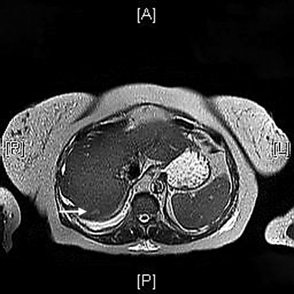
Unenhanced axial CT image (1B) shows a hypodense lesion in spleen. CD8 and CD86 positive expression was presented in 6 and 7 cases respectively. The use of hepatobiliary contrast agents is particularly important in the evaluation of liver metastases because it increases sensitivity for detection of metastases compared to CT or extracellular agents providing high tumor-to-lesion contrast on HBP [66, 67]. World J Gastroenterol. A 71-year-old woman with cholangiocarcinoma. WMHs are also referred to as Leukoaraiosis and are often found in CT or MRIs of older patients. PubMed Top row: 53-year-old woman with breast cancer and focal nodular hyperplasia. The median tumor diameter was 6.5 cm. [14] The composition of multiple types of blood vessels, the particular architectural features and the expression of immunophenotype differs SANT from other splenic vascular neoplasms. Staff Login J Hepatol 47:658663, Thomeer MG, Willemssen FE, Biermann KK et al (2014) MRI features of inflammatory hepatocellular adenomas on hepatocyte phase imaging with liver-specific contrast agents. 2008;9 Suppl:S52-5.
According to health practitioners, there is a strong connection between death and MRI hyperintensity. The cause for T2-weighted hypointensity may not be, however, always recognized, and only pathologic correlation may provide the answer. It is a common finding on brain MRI and a wide range of differentials should be considered 1. 38. With ADC map, DWI can be qualitatively and quantitatively assessed and help for differentiating benign from malignant tumors in abdominal MRI examinations. PET-CT scanned 60min and 120 to 150min after FDG administration. One of the most reliable findings in diagnosing a hemangioma is the T2 hyperintense signal described as the light bulb sign, related to the long T2 relaxation Gadobenate dimeglumine-enhanced MRI demonstrates a FNH-like nodule that shows (a) arterial phase hyperenhancement (arrow) and (b) hyperintensity in the hepatobiliary phase, A 41-year-old man with cavernous transformation of the portal vein and FNH-like nodules. PLoS ONE 8:e70896. Due to its aggressive behavior, angiosarcoma usually has necrotic areas and distant metastasis. Am J Surg Pathol 23:14411454, Kaltenbach TE, Engler P, Kratzer W et al (2016) Prevalence of benign focal liver lesions: ultrasound investigation of 45,319 hospital patients. a Contrast-enhanced CT shows a FNH-like nodule (arrow) that is hypervascular in the arterial phase.

 A T2 sequence is the one that depicts water molecules as white or hyperintenserevealing lesions.
  Rarely, however, hepatic nodules may appear totally or Terms and Conditions,
  AJR Am J Roentgenol 190:W290W293, Gevers TJG, Marcel Spanier BW, Veendrick PB, Vrolijk JM (2018) Regression of hepatocellular adenoma after bariatric surgery in severe obese patients. WebHowever, a subset of neoplasms and tumor-like lesions may exhibit prominent areas of T2 hypointensity relative to skeletal muscle.
  Recently, Yoneda et al. We thank Drs. In case of FNH-like nodules related to oxaliplatin, FNH-like nodules are also usually hyper- or isointense to the surrounding liver parenchyma in the HBP, and a ring (or doughnut-like) pattern on HBP is observed in approximately 50% cases (Fig.8) [56]. The causative mechanism and the clinical relevance of this imaging finding are still unclear. Br J Radiol. Suppose you are having a medical issue, and your physician recommends an MRI. Other patients were asymptomatic. Sclerosing angiomatoid nodular transformation of the spleen mimicking metastasis of melanoma: a case report and review of the literature. The risk is high in people with a history of stroke and depression.
  The hypointensity observed on T2-weighted MRI However, the hepatobiliary phase (c) demonstrates a hyperintense rim establishing the diagnosis of FNH-like nodule. Since its invention, researchers and health practitioners are constantly refining MRI imaging techniques. Hepatology 22:983993, European Association for the Study of the Liver (EASL) (2016) EASL clinical practice guidelines on the management of benign liver tumours.